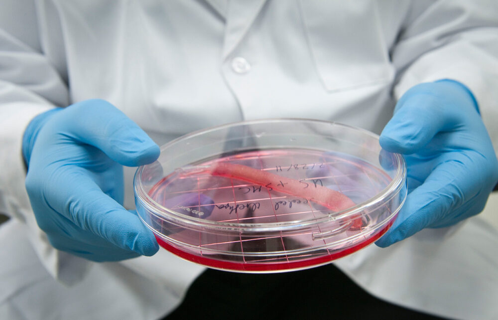
Researchers from Vienna’s Austrian Academy of Sciences have grown a bundle of human stem cells into a tiny artificial “heart” the size of a sesame seed.
The pulsating mass is the first self-organizing miniature organ to resemble the human heart, including a hollow chamber enclosed by a wall of cardiac-like tissue.
Significantly, unlike previous versions of these tiny heart organs (called cardioids), the scientists didn’t use artificial scaffolding to bind the cells together. Instead, the cells organised themselves to grow a hollow chamber.
Other studies have managed to grow self-organizing eye organoids, self-organizing brain organoids, and self-organizing gut organoids using similar signaling techniques.
“It’s not that we are using something different than other researchers, but we are just using all of the signals known,” explains biologist Sasha Mendjan from the Austrian Academy of Sciences in Vienna.
By creating more lifelike heart models, scientists are hoping to gain a better understanding of how the cardiac system responds to disease.
After just one week of growing in the lab, researchers noticed their mass of cells had formed a 3D structure that could beat rhythmically, squeezing liquid in and out of its chamber-like cavity.
To create the organoids whose cells self-organise like those in an embryo, the authors of the new study programmed human pluripotent stem cells, which have the ability to differentiate into any kind of tissue, into various types of cardiac cells. They aimed to create the three tissue layers present in the walls of a heart chamber, one of the first parts of the organ to develop. Next, the researchers immersed the stem cells in different concentrations of growth-promoting nutrients until they found a recipe that coaxed the cells to form tissues in the same order and shape seen in embryos.
After 1 week of development, the organoids are structurally equivalent to the heart of a 25-day-old embryo. At this stage, the heart has only one chamber, which will become the left ventricle of the mature heart. The organoids are about 2 millimeters in diameter and include the main types of cells typically present in this stage of development: cardiomyocytes, epithelial cells, fibroblasts, and epicardium. They also have a clearly defined chamber that beats at 60 to 100 times per minute, the same rate of an embryo’s heart around the same age.
The minihearts, which have so far survived for more than 3 months in the lab, will help scientists see heart development in unprecedented detail. They might also reveal the origins of cardiac problems like congenital heart defects in babies and cardiac cell death after heart attacks.


