Doctors at Postgraduate Institute of Medical Education and Research (PGIMER), Chandigarh, have created history by successfully removing a large brain tumour through the nose of a one year and four-months-old girl child.
She is said to be the youngest patient in the world to have such an endoscopic surgery.
A team of endoscopic skull base surgeons, Dr Dhandapani SS and Dr Sushant, both from Department of Neurosurgery, and Dr Rijuneeta from Department of ENT operated upon child hailing from Uttarakhand. She was referred to PGIMER with complaints of loss of vision.
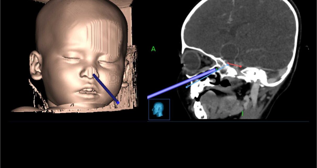
As per the doctors, the child was normal and playful following visual stimuli a few months back. For the last 20 days, the mother noticed that the child was not following anything shown to her. The child’s MRI revealed a calcified brain tumour at the base of the skull suggestive of craniopharyngioma of size three cm, large for a child of one year, close to critical neural structures such as optic nerves and hypothalamus.
The tumours are usually operated on through open surgery, and the remaining part is treated with radiation therapy. Over the last few years, such tumours are being removed through the nose endoscopically by neurosurgeons teaming with ENT surgeons among patients older than six years.
However, endoscopic removal through the nose is highly challenging in small children because of small nostrils, immature bones at the skull base, and proximity to crucial blood vessels.
Despite the challenge, Dr Dhandapani chose the endonasal corridor, as the skull opening and brain retraction are avoided if operated through the nose.
Reaching the brain tumour was difficult, as the bones and sinuses were immature. The typical air sinus, which usually gives a corridor to reach up to the tumour base, was absent in this child.
The nasal stage was performed by ENT surgeon Rijuneeta, while the skull base part was completed by Dr Dhandapani and Dr Sushant.
Extensive drilling of the immature bones with a diamond drill was carried out using computer navigation to create a tumour removal corridor.
Doctors say the brain tumour was dissected from critical structures using angled endoscopes and removed through the nose despite very little working space.
As endonasal endoscopic surgery of brain tumour can cause brain fluid leakage through the nose, the vascularized flap taken from inside the nose was used to seal the operative corridor along with fascia and glue. After a six-hour long surgery, the child was kept in the ICU and recovered very well. Ten days after the surgery, the child is doing great with improved vision and no complications, with a CT scan showing almost complete removal.